Clinical pilot NEW
CBCT + IOS fusion: every tooth with its real root, the scanned gingiva, and the bone
One planning model from a CBCT volume and the intraoral scans. The crowns come from the intraoral scan, the roots and the bone from the CBCT, and each tooth is delivered as a closed solid you can move, extract or plan against in exocad.
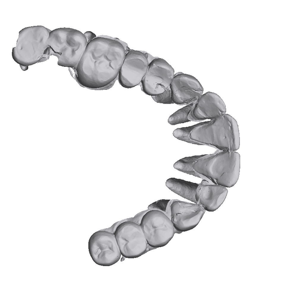
What you get
How it works
- Segment the CBCT. An open neural network trained for dental CBCT (DentalSegmentator, nnU-Net) labels the upper and lower teeth and the two jaws in the volume.
- Register the two modalities. Only the crown part of the CBCT teeth, the part above the bone that both scans see, is fitted to the intraoral scan, so gum and roots cannot pull the alignment.
- Split the teeth. Each crown on the intraoral scan seeds its own tooth in the CBCT; neighbouring teeth separate at their contact instead of being cut by hand.
- Fuse. The intraoral scan is cut into crowns and gingiva along the gum line, the CBCT root is taken below each crown, and the two are joined into one closed solid per tooth.
Every step is measured against a real case prepared by a technician, and the measurements, including the parts that are not there yet, are written up on the research page.
Measured on the pilot case
| What | Result |
|---|---|
| Teeth surface against the technician's reference | 0.2 mm at the median, 0.56 mm at the 90th percentile |
| CBCT to intraoral registration | 0.34 mm residual, 84 % of crown points within 1 mm |
| Gum-line cut against the technician's cut | intersection over union 0.89 |
| Teeth found | 14 of 14 on the upper arch, each as its own solid |
| Time per case | about 10 minutes per arch on a workstation, including the CBCT segmentation |
What is still being worked on. Some anterior teeth are not yet consistent closed solids where the intraoral scan folds over itself on the palatal side; crowns that touch are joined by a short strip rather than cut along the contact; metal restorations bloom in the CBCT and need a dedicated rule; and the bone layer is cropped tighter than a technician would. These are the reasons the product is a pilot and not a button yet.
Scans you need
- CBCT as DICOM: a folder of slices or one multi-frame file. Voxel size of 0.15 to 0.3 mm. A scan with the arches slightly apart segments more cleanly than one in occlusion.
- Intraoral scan of each arch you want, as binary STL, the same files you would send to a lab.
- Both taken close in time. The pilot case had seven months between them and still registered, but the gum line moves.
Frequently asked questions
What does the fusion model contain?
Four layers as STL: every tooth as one closed solid made of the intraoral-scan crown and the CBCT root, the gingiva from the intraoral scan with the crowns cut out at the gum line, the alveolar bone from the CBCT, and all three together. Each tooth is also provided as its own file.
Which scans are needed?
One CBCT as DICOM (a folder of slices or a single multi-frame file) and an intraoral scan of each arch you want as binary STL. The two should be taken close in time, and a CBCT with the arches slightly apart segments more cleanly than one in occlusion.
How accurate is the result?
On the pilot case the crowns and roots lie within 0.2 mm of a technician's hand-made reference at the median, the registration between CBCT and intraoral scan converges to 0.34 mm with 84 percent inliers, and the gum-line cut agrees with the technician's at an intersection-over-union of 0.89. The remaining differences are documented openly.
How is this different from a VeX model?
A VeX model is built from two intraoral scans and keeps only the root pockets the teeth scan captured. The fusion model adds the CBCT, so each tooth carries its complete root inside the bone and the bone itself is part of the model.
Is it available now?
It is in a clinical pilot. Cases are run with the clinic on request and returned as STL; leave your email below and we will come back to you.
Run a case with us.
Leave a work email. We will get in touch about sending a CBCT and the intraoral scans and return the fused model as STL.